Manufacture
of MacroPorous *
Calcium Hydroxyapatite and
Tri-Calcium Phosphate
Bioceramics
N. Ozgur ENGIN and A. Cuneyt TAS
Dept. of Metallurgical and
* N. O. Engin
and A. C. Tas, "Manufacture of Macroporous Calcium Hydroxyapatite
Bioceramics," Journal of The European Ceramic
Society, Vol. 19 (13-14), 2569-2572 (1999).
(--> download pdf)
* N. O. Engin
and A. C. Tas, ”Preparation of Porous
Ca10(PO4)6(OH)2 and Beta-Ca3(PO4)2 Bioceramics,”
Journal of The American Ceramic Society, 83(7), 1581-1584 (2000).
(--> download: ha-tcp-porous.pdf)
* N. O. Engin
and A. C. Tas, "Manufacture of Macroporous Calcium Hydroxyapatite
Bioceramics,"
100th Annual Meeting of the American Ceramic Society,
May 3-8, 1998, Cincinnati, Ohio, USA, Oral Presentation.
* N. O. Engin
and A. C. Tas, "Manufacture of Macroporous HA Bioceramics,"
New Developments in High-Temperature Ceramics Conference
(Sponsored by Office of Naval Research (
* N. O. Engin
and A. C. Tas, "Manufacture of Macroporous Calcium Hydroxyapatite
Bioceramics,"
IV. Ceramics Congress,
* N. O. Engin,
"Manufacture of Macroporous Calcium Hydroxyapatite (HA) and Tr-Calcium
Phosphate (TCP) Bioceramics,"
M.Sc. Thesis, January 1999 (Supervisor:
Dr. A.C. Tas) .
* Patent No: TR-9900038,
Trabecular bones of almost
all vertebrate organisms do basically consist of macroporous
(55 to 70% interconnected porosity) bone mineral, i.e., calcium hydroxyapatite (HA: Ca10(PO4)6(OH)2). The macroporosity observed in the trabecular
bones then allows the ingrowth of the soft tissues
and organic cells into the bone matrix.
Sub-micron,
chemically uniform, and high phase-purity HA (or TCP) powders produced in our
laboratory were mixed, under vigorous ultrasonification,
with either methyl cellulose or polyethyleneimine of
appropriate amounts in the form of an aqueous slurry of proper viscosity and
thickness. The ceramic cakes produced in this way were then carefully dried in
an oven in the temperature range of 50 to 90°C. Dried cakes of porous HA (or
TCP) were then physically cut into various prismatic shapes. These parts were then
slowly heated in an air atmosphere to the maximum sintering temperature of
1250°C. The HA (or TCP) bioceramic parts obtained by
this “foaming technique” were found to have tractable and
controllable interconnected porosity in the range of 60 to 90%, with typical
average pore sizes ranging from 100 to 250 microns. Sample characterization was
mainly achieved by SEM (scanning electron microscopy) studies and three-point
bending tests.
INTRODUCTION
With
the growing demands of bioactive materials for orthopaedic
as well as maxillofacial surgery, the utilization of calcium hydroxyapatite (HA, with Ca/P = 1.667) and tricalcium phosphate (TCP, with Ca/P = 1.50) as fillers,
spacers, and bone graft substitutes has received great attention mainly during
the past two decades, primarily because of their biocompatibility, bioactivity,
and osteoconduction characteristics with respect to
host tissue (1-3).
For
certain periods, attention was particularly placed on the fabrication of bioceramics with “porous” configuration because
the porous network allows the tissue to infiltrate, which further enhances the
implant-tissue attachment (4-13). In a porous form, hydroxyapatite
ceramics can be colonized by bone tissue with the same characteristics as peri-implanted tissues (14). For colonization of the pores
to take place, they must be larger than 50-100 µm (13) or even 250-300 µm
according to some researchers (15-17).
EXPERIMENTAL PROCEDURE
The
hydroxyapatite (and tri-calcium phosphate) powders
produced in our laboratory (18), with average particle size of 0.6-0.7 µm, were
used (19) to prepare an HA (or TCP) slurry essentially consisting of methyl
cellulose to form spongy bioceramic cakes and bodies
of differing porosity simulating that typical of bone. Solutions containing the
HA (or TCP) powders and polymeric agents were treated with an ultrasonic
disruptor (Misonix, Inc., Model: XL2015, NY,
Scanning
electron microscopy (SEM, Jeol Corp., Model:
JSM-6400,
RESULTS
The
novel foaming method used in this study (19-20), to produce macroporous
calcium hydroxyapatite bioceramic
parts, were shown to be successful in the attainment of relative porosity over
the range of 60 to 90%. The control of porosity in the HA samples were found to
be achieved by essentially changing the amount of polymeric agent used in the
slurries.
The
pore sizes in our HA (or TCP) bioceramics were
typically distributed in the range of 100 to 400 µm. The pores were
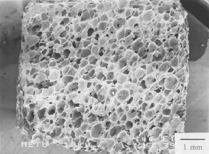
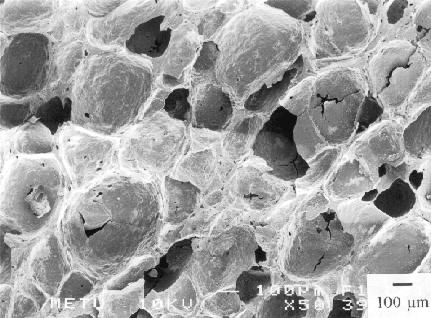
interconnected. The SEM micrographs given in Figures 1 to 3 display the
microstructures of macroporous HA parts produced in
our laboratory with 60, 75, and 90% relative porosity, respectively.
This technique of porous ceramic manufacture may
easily be used in other ceramic phases and materials, and therefore, has a vast
potential for future technological applications.
Click on the pics to display
Figs. 1a & 1b
SEM micrographs of 60% porosity HA (or TCP) bioceramic
parts
Click on the pics to display
Figs 2a
& 2b SEM micrographs of 75% porosity HA bioceramic parts


Figs 3a
& 3b SEM micrographs of 90% porosity HA bioceramic parts


Click on the pics to display
Acknowledgments
This
study has been supported by the research project of TÜBITAK / Misag-58. The
authors are also thankful to the staff and researchers of the Department of
Petroleum and Natural Gas Engineering (METU) for performing the mercury porosimetry and computerized tomography analysis.
REFERENCES
1.
K. de Groot, “Bioceramics
Consisting of Calcium Phosphate Salts,” Biomaterials, 1, 47-50 (1980).
2. M. Jarcho, “Calcium Phosphate
Ceramics as Hard Tissue Prosthetics,” Clin. Orthop. Rel. Res., 157, 259
(1981).
3. C. J. Damien and J. R. Parsons, “Bone Graft and Bone Graft
Substitutes: A Review of Current Technology and Applications,” J. Appl. Biomaterials, 2, 187-208 (1990).
4. E. W. White and E. C. Shors,
“Biomaterial Aspects of Interpore 200 Porous Hydroxyapatite,” Dental Clin.
N. Am., 30, 49, (1986).
5. R. E. Holmes, V. Mooney, R. Bucholz, and A.
Tencer, “A Coralline Hydroxya
patite Bone Graft Substitute,” Clin. Orthop. Rel.
Res., 188, 252-262, (1984).
6. R. E. Holmes, “Bone Regeneration within a Coralline Hydroxyapatite Implant,” Plast.
Reconstr. Surg., 63, 626
(1979).
7. E. C. Shors, E. W. White, and G. Kopchok, “Biocompatibility, Osteoconduction
and Biodegradation of Porous Hydroxyapatite and
Calcium Carbonate in Rabbit Bone Defects,” Mater. Res. Soc. Symp. Proc., 110, 211-217 (1989).
8. H. Ohgushi, M. Okumura, and T. Yoshikawa,
“Bone Formation Process in Porous Calcium Carbonate and Hydroxyapatite,” J. Biomed.
Mater. Res., 26, 885-895 (1992).
9. Dean-Mo Liu, “Fabrication and Characterization of Porous Hydroxyapatite Granules,” Biomaterials, 17, 1955-1957
(1996).
10. C. Klein, K. de Groot, C. Weiqun, L. Yubao, and Z. Xingdong, “Osseous Substance Formation Induced in
Porous Calcium Phosphate Ceramics in Soft Tissues,” Biomaterials, 15,
31-34 (1994).
11. M. Fabbri, G. C. Celotti,
and A. Ravaglioli, “Hydroxyapatite-based
Porous Aggregates: Physico-chemical Nature,
Structure, Texture and Architecture,” Biomaterials, 16, 225-228 (1995).
12. M. Fabbri, G. C. Celotti,
and A. Ravaglioli, “Granulates Based on Calcium
Phosphate with Controlled Morphology and Porosity for Medical Applications: Physico-chemical Parameters and Production
Technique,” Biomaterials, 15, 474-477 (1994).
13. J. C. Le Huec, T. Schaeverbeke,
D. Clement, J. Faber, and A. Le Rebeller,
“Influence of Porosity on the Mechanical Resistance of Hydroxyapatite Ceramics under Compressive Stress,”
Biomaterials, 16, 113-118 (1995).
14. N. Passuti, G. Daculsi,
J. M. Rogez, S. Martin, and J. V. Bainvel,
“Macroporous Calcium Phosphate Ceramics
Performance in Human Spine Fusion,” Clin. Orthoped.,
248, 169-176 (1989).
15.
J. J. Klawiter, J. G. Bagwell, A. M. Weinstein, B. W. Sauer, and J. R. Pruitt,
“An Evaluation of Bone Growth into Porous High Density
Polyethylene,” J. Biomed. Mater. Res., 10,
311-321 (1976).
16. P. S. Eggli, W. Müller,
and R. K. Schenk, “Porous Hydroxyapatite and Tricalcium Phosphate Cylinders with Two Different Pore Size
Ranges Implanted in the Cancellous Bone of
Rabbits,” Clin. Orthoped.,
232, 127-138 (1987).
17. G. Daculsi and
18. A. C. Tas, “Synthesis of the Two
Inorganic Phases (HA: Ca10(PO4)6(OH)2 and TCP: Ca3(PO4)2) of Synthetic Bone
Applications by A Chemical Precipitation Method,” Patent No: TR 1995
01422 B, Turkish Patent Institute,
19. N. Ö. Engin, “Manufacture of Macroporous Hydroxyapatite Bioceramics,” M.Sc. Thesis,
METU, January 1999 (Thesis supervisor: Dr. A. Cüneyt
Tas)
20. A. C. Tas (Inventor), “Manufacture
of Macroporous Calcium Hydroxyapatite
(HA) and Tri-calcium Phosphate (TCP) Bioceramics,”
Patent No: TR-9900038 (Owner: Turkish Scientific and Technical Research
Organization, TUBITAK), Turkish Patent Institute,